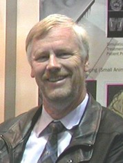
Dr. Horst Bruning is Adding New Dimensions to Medical Imaging
Dr. Horst Bruning is Adding New Dimensions to Medical Imaging

The place where science and technology meet is comfortable ground for Dr. Horst Bruning, president and CEO of Exxim Computing Corporation. Dr. Bruning, whose PhD is in physics, first became interested in the subject during a year as a high school exchange student in New York.
"There was a very good physics teacher there. I liked the guy and he made it exciting to look into physics. It explains it all, right? These physics books, like A Short History of Nearly Everything, they're popular books, and they're basically the physicist's view of the world - things which you observe, and you try to explain them using the language of math, so it's reproducible.
"Our scientific approach through observations, that's what I kind of liked, because it makes you feel like you have a solid foundation - you can come back to it and it will be reproducible every time. Today, the scientific methodology has been expanded into genetics and these modern bioscience/life science industries. But physics is kind of at the basis of it."
After completing an undergraduate degree in his native Germany, he worked at CERN in Geneva, Switzerland, the world's largest particle physics laboratory. He supported the lab's quest to discover new subatomic particles. "There was a time there in the 70s where there were lots of new particles coming up, like every month a new one, and the experimental work that we did there had to do with particle detectors. I was an experimental physicist building these detectors to find pi mesons and psi bosons and things like that, calculating their energy and their charge and these things."
He earned his PhD then and decided to apply this technology to "something useful," as he puts it. "Maybe particle physics is useful in the long run, but not directly." He took a job at Siemens Central Research and Siemens Medical, building x-ray detectors and getting into the more computer-driven world of computer tomography (the building of images detected by x-rays, ultrasound, or other means using computers).
"I used the physics background to go into electrical engineering and software and imaging applications, but other physicists stay in the scientific realm and do cosmology or things like that. The mathematics is very similar but I am more applications-driven; I want to build machines which are out there in the field and help people and can be sold and turned into a business. I'm an applied physicist."
He contributed to computed tomography (CT) scanners (originally known as CAT scanners, an acronym for computed axial tomography) at Siemens and at Imatron in South San Francisco. Aside from finding that he enjoyed his work, he made another important discovery: he wanted to live in the Bay Area. "I went back to Germany and kind of didn't like it anymore - too much rain! California is like a diode - people come here but they hate to go the other way."
He returned to the area to work at Invision, a security scanner company that was just recently bought out by GE, and continued his work on CT technology and visualization at TeraRecon in San Mateo before founding Exxim - the name comes from "extreme x-ray imaging for the XXI century" - in 2002.
"You see opportunities and when you are in a company, you try to do it within the company, obviously, but sometimes it's not possible because it doesn't fit the business plan of that particular company. If you feel strong enough about it and you really want to do it, there's only one way: to do it yourself and start a new company. I think this is how many of the new small companies here in the area have come up. People had an idea, wanted to realize it, couldn't do it in the environment they were in, and started by themselves. There are lots and lots of very small companies like this, five-person companies that do really interesting high-tech stuff."
There's no shortage of really interesting stuff going on at Exxim. While CT scanners typically take two-dimensional images of a cross- section of a person, animal, or object - usually referred to as a "slice" - the latest scanners use a wide area detector to scan an entire three-dimensional volume, which has been given the name "cone- beam tomography." Exxim's primary product in this area is COBRA, software that takes the raw data output by a cone-beam CT scanner and reconstructs it into a "volume output image," which is essentially a navigable, color 3-D image.
"We've found some very interesting applications for this. One application was almost unexpected for us - it's in dental x-ray imaging. Dental offices use a lot of x-rays and normally you have these little films that you stick in your mouth. Today, they have these small digital reception device detectors that you can stick in there, but basically you get one bite wing, as they call it, radiograph. Now you can have an area detector x-ray so as you go around the head of the patient, you get a three-dimensional image of the jaws and the teeth. The images are fantastic.
"People use this imaging technique to assess the quality of the jaw bone for an implant. Many people get implants today as opposed to a bridge or something to replace lost teeth and the bone needs to have a certain quality - they drill a metal screw in the bone, so the bone needs to be strong enough to withstand that, and also you don't want to screw into the nerve and do damage. So you can assess the whole geometry of the jaws and the facial bones. That's an enormously fast- growing market, and these people use our software."
Exxim's technology is also being used in scanning small animals, such as mice, which are used in research settings at pharmaceutical institutes and universities. "You can scan the volume of the mouse rapidly with these volume scanners." This allows researchers to follow internal changes in the animal's anatomy, which is useful in research in research for new treatments for tumors, among other things.
Another new application for Exxim's technology is a security application for detecting bombs in luggage. "In one single scan, you can scan a whole bag in volume and find hidden explosives in there. There's still some work to be done, though."
Dr. Bruning also continues to utilize his expertise in building detectors in a project he is working on with Real-Time Radiography, an Israeli company out of the Jerusalem Technology Park which is making a new material to convert the incoming x-rays into electrical signals more efficiently than it's done today. While current technology relies on light generated by the x-rays interaction with a compound like cesium oxide, the new technology uses mercuric iodide, a heavy compound which generates electrical charges when exposed to x- rays rather than light. This allows for images with greater sensitivity and detail. "It gives you a bigger signal and you can make the images using a lower dose of x-rays." A product using Exxim's detector developed in conjunction with Real-Time Radiography is expected to be on the market about two years from now.
Also in this issue...
- ZANTAZ Launches First Archive On Demand
- Javelin Strategy & Research Aiming High and Succeeding
- Business Bits
- Executive Profile: Dr. Horst Bruning is Adding New Dimensions to Medical Imaging
- SFG Bancorp Expanding with Online Mortgage Lending Application
- West Valley Staffing Group Growing With Its Clients
- Springtime is the Perfect Time to Enjoy Pleasanton's Parks
- Tri-Valley One-Stop Offers Help with Hiring, On-the-Job Training Incentives
- It's Easy Being Green with Expert Assistance from Alameda County's StopWaste Program
- Open Heart Kitchen Provides Nutritious Meals to Area Children, Families, Seniors
- Hacienda Index
- Calendar


